TMD Referral Center
for
Medical Professionals

Objective Assessment of Jaw Function
What we don’t measure, we can only assume. What we measure, we can understand.
When Anatomy Isn’t the Whole Story
Some aspects of jaw function cannot be fully understood from a clinical examination or conventional dental imaging alone.
Our diagnostic approach incorporates objective functional measurements to examine how the masticatory system is working. Depending on the clinical question, we can evaluate mandibular movement, masticatory muscle activity, and TMJ function, providing information that complements the clinical examination and imaging findings.
Masticatory Muscle Electromyography (EMG) -Objective Assessment of Masticatory Muscle Activity

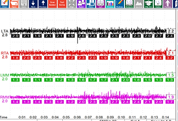
Electromyography (EMG) provides an objective way to record this muscle activity. Using surface electrodes placed over selected masticatory muscles, we can record and analyze their electrical activity in real time. This allows us to evaluate muscle function under different conditions rather than relying solely on what can be observed during a clinical examination.
EMG can provide information about the level and pattern of muscle activation, including differences in activity between the right and left sides, changes in muscle recruitment, and how the muscles respond during different mandibular tasks.
For example, a patient may appear to have relatively normal mandibular movement during a routine examination, while functional recording may reveal differences in muscular recruitment or an uneven pattern of activity. Conversely, symptoms may be present even when the clinical examination does not reveal an obvious structural abnormality.
The ability to observe muscle activity during different functional conditions is particularly valuable because the masticatory system does not operate through isolated muscles. The muscles work as a coordinated system to position and stabilize the mandible. Changes in one part of this system can influence how other muscles participate in mandibular function.
Our EMG assessment can therefore be used as one component of a broader functional evaluation. The recorded muscle activity can be considered together with mandibular movement, TMJ findings, occlusal contacts, and the patient's clinical presentation.
The value of EMG is not simply in obtaining a measurement, but in adding objective functional information to the diagnostic picture. It allows us to document muscular behavior, establish a functional baseline, and compare findings when appropriate during subsequent stages of evaluation or treatment.
Selected Literature:
Szyszka-Sommerfeld L, et al. (2023). Surface electromyography in the assessment of masticatory muscle activity in patients with pain-related temporomandibular disorders: a systematic review. Frontiers in Neurology, 14, 1184036.
https://www.frontiersin.org/journals/neurology/articles/10.3389/fneur.2023.1184036/full
Dorosz T, Mańko A, Ginszt M. (2024). Use of Surface Electromyography to Evaluate Effects of Therapeutic Methods on Masticatory Muscle Activity in Patients with Temporomandibular Disorders: A Narrative Review. Journal of Clinical Medicine, 13(3), 920.
DOI: 10.3390/jcm13030920.
https://www.mdpi.com/2077-0383/13/3/920
Hugger S, Schindler HJ, Kordass B, Hugger A. (2013). Surface EMG of the masticatory muscles (Part 3): Impact of changes to the dynamic occlusion. International Journal of Computerized Dentistry, 16(2), 119–123.
This is especially relevant to your clinical concept, because it specifically reviews masticatory sEMG in relation to changes in dynamic occlusion, rather than discussing EMG in isolation.
The K7x CMS Computerized Mandibular Scanning (Jaw Tracking)
The mandible is not a static structure. Every time we speak, chew, swallow, or open and close the mouth, the jaw follows a specific pattern of movement that reflects the coordinated function of the temporomandibular joints, muscles, teeth, and neuromuscular system.
A clinical examination allows us to observe many aspects of this function, but it does not necessarily show us the precise path the mandible follows during movement.
Mandibular kinematic analysis provides an objective way to record and visualize these movements. Using a computerized tracking system, we can capture the trajectory of the mandible in three dimensions and examine how the movement develops throughout opening, closing, and other functional movements.
This can provide information about movement symmetry, coordination, deviations, restrictions, and changes in the trajectory that may not be apparent during a routine examination.
The purpose is not simply to record how far the jaw can move. The movement itself provides functional information — how the mandible travels through space, how consistently it follows its path, and how the movement relates to the overall function of the masticatory system.
Because the recording system is lightweight and designed to allow normal access to the patient’s mouth, measurements can be obtained without substantially interfering with the movements being evaluated.

Selected Literature:
Woodford SC, Robinson DL, Mehl A, Lee PVS, Ackland DC. (2020). Measurement of normal and pathological mandibular and temporomandibular joint kinematics: A systematic review. Journal of Biomechanics, 111, 109994.
DOI: 10.1016/j.jbiomech.2020.109994.
https://www.sciencedirect.com/science/article/abs/pii/S0021929020304176
D’Amico C, et al. (2022). Efficacy of Kinematic Parameters for Assessment of Temporomandibular Joint Function and Dysfunction: A Systematic Review and Meta-Analysis. Bioengineering, 9(7), 269.
DOI: 10.3390/bioengineering9070269.
https://www.mdpi.com/2306-5354/9/7/269
Zhang R, Lai D, Zhang F, et al. (2025). A Comparative Analysis of Temporomandibular Disorders Using a Jaw Motion Analyzer and Surface Electromyography. International Dental Journal, 75(3), 1843–1853.
https://www.sciencedirect.com/science/article/pii/S0020653925001030?via%3Dihub
K7x ESG Electrosonography (TMJ Vibration Evaluationi System)

The temporomandibular joints are dynamic structures. During opening and closing, the mandibular condyle, articular disk, and surrounding joint structures move together through a coordinated functional pathway. When this movement is altered, mechanical events within the joint may produce vibrations, clicks, or other detectable sounds.
Computerized joint sonography records vibrations from the right and left TMJs continuously while the jaw is moving. This differs from imaging studies such as CBCT or MRI, which provide detailed anatomical information captured at a particular jaw position or at selected positions during an examination.
Rather than simply determining whether a joint makes a sound, sonography allows us to examine when during opening or closing the vibration occurs, its relative intensity and frequency characteristics, which joint it originates from, and whether the event is consistently reproduced during repeated movements.
When sonography is evaluated together with computerized mandibular tracking, the timing of a vibration can also be correlated with the phase and position of mandibular movement at which it occurs. This provides additional objective information about joint mechanics and disk-condyle coordination and may help identify functional patterns that warrant further clinical evaluation or imaging.
Sonography does not directly visualize the articular disk and is not a replacement for MRI, CBCT, or a comprehensive clinical examination. Instead, these technologies provide complementary information: imaging helps evaluate joint anatomy, while sonography helps evaluate how the joint behaves dynamically during function.
Selected Literature:
Sharma S, Crow HC, McCall WD Jr, Gonzalez YM. (2013). Systematic Review of Reliability and Diagnostic Validity of Joint Vibration Analysis for Diagnosis of Temporomandibular Disorders. Journal of Oral & Facial Pain and Headache, 27(1), 51–60.
DOI: 10.11607/jop.972
https://www.jofph.com/articles/10.11607/jop.972
Sharma S, Crow HC, Kartha K, McCall WD Jr, Gonzalez YM. (2017). Reliability and diagnostic validity of a joint vibration analysis device. BMC Oral Health, 17, 56.
DOI: 10.1186/s12903-017-0346-9
https://link.springer.com/article/10.1186/s12903-017-0346-9
Garcia AR, Madeira MC, Paiva G, Olivieri KA. Effect of occlusion on joint sounds in asymptomatic individuals. Effect of occlusion on joint sounds in asymptomatic individuals.
T - Scan Novus - Digital Occlusal Mapping — Measuring the Dynamics of Occlusion
The way the teeth come into contact is an important part of masticatory system health. An uneven distribution of occlusal contacts can create excessive forces in particular areas and may contribute to increased masticatory muscle activity as the neuromuscular system works to maintain mandibular stability and postural balance.
Identifying where these excessive occlusal forces are concentrated can therefore provide valuable information when trying to understand the source of muscular tension.
Digital occlusal mapping with the T-Scan provides information that conventional occlusal paper cannot. Rather than showing only where the teeth have contacted, it allows us to observe the entire dynamic sequence of occlusion — from the very first tooth contact through the progression to maximum intercuspation.
We can see where the mandible is positioned at the initial point of contact, how long it remains at that initial contact, and how the distribution of occlusal forces changes as the jaw continues into full occlusion. At the same time, we can evaluate where the center of occlusal force is concentrated and how it moves throughout the complete occlusal cycle.
The system also provides a quantitative representation of the relative contribution of individual occluding tooth pairs. We can see what percentage of the overall occlusal force is being carried by different areas of the dentition and how that distribution changes during function.
This dynamic information provides a much more detailed picture of the patient's occlusion than conventional articulating paper alone. Instead of simply identifying contact points, we can evaluate where the force is concentrated, when it occurs, how it develops, and how it shifts as the mandible moves into full occlusion. For us, this information is particularly valuable when evaluating the relationship between occlusion and masticatory muscle activity. It can help identify areas of excessive occlusal loading and provide objective guidance when determining the direction of occlusal adjustment as part of a broader process of functional occlusal equilibration and muscular rebalancing.
Selected Literature:
Chowdhary R, Sonnahalli NK. (2024). Clinical applications of the T-Scan quantitative digital occlusal analysis technology: a systematic review. International Journal of Computerized Dentistry, 27(1), 49–86.
DOI: 10.3290/j.ijcd.b3945153
Cerna M, Ferreira R, Zaror C, Navarro P, Sandoval P. (2015). Validity and reliability of the T-Scan III for measuring force under laboratory conditions. Journal of Oral Rehabilitation, 42(7), 544–551.
DOI: 10.1111/joor.12284
https://onlinelibrary.wiley.com/doi/abs/10.1111/joor.12284
Seth-Johansen C, Gotfredsen K. (2025). Validity and reliability of digital occlusal analyzing methods in Dentistry: A systematic review. Journal of Dentistry, 163, 106124.
DOI: 10.1016/j.jdent.2025.106124
https://www.sciencedirect.com/science/article/abs/pii/S0300571225005706

Correlating Occlusion and Muscle Activity
Because T-Scan and surface EMG provide different types of functional information, recordings can be evaluated together to examine the relationship between tooth contact patterns and masticatory muscle activity.
A recording obtained with the patient's existing occlusion can document the distribution and timing of tooth contacts while EMG simultaneously evaluates the activity of the monitored muscles. A second recording obtained with a properly adjusted occlusal orthotic provides an opportunity to observe how the muscle activity pattern compares under a different occlusal condition.
The purpose of this comparison is not to assume that occlusion is the cause of a particular muscle pattern. Rather, it allows us to objectively examine whether changes in occlusal contact coincide with measurable changes in muscular activity, symmetry, recruitment, or relaxation.
When a reproducible difference is observed, it becomes another piece of functional information that can be considered together with the clinical examination, symptoms, mandibular movement, joint findings, and other diagnostic information.
existing habitual occlusion
T-Scan image + corresponding EMG recording
.png)
.png)
occlusal neuromuscular orthotic
T-Scan image + corresponding EMG recording (after first one week adjustment)
.png)
.png)
Selected Literature:
Wieczorek A, Loster J, Loster BW, Sierpińska T, Gołębiewska M. (2013). Relationship between Occlusal Force Distribution and the Activity of Masseter and Anterior Temporalis Muscles in Asymptomatic Young Adults. BioMed Research International.
https://pmc.ncbi.nlm.nih.gov/articles/PMC3591202/
Tecco S, et al. (2015). Activity of the masticatory muscles and occlusal contacts in young adults with and without orthodontic treatment. BMC Oral Health.
https://link.springer.com/article/10.1186/s12903-015-0099-2
Comparative evaluation of night guard and stabilization splint therapy on temporomandibular joint function using surface electromyography and digital occlusal analysis: a DC/TMD-based study. BMC Oral Health (2026). It compared stabilization splints, night guards, and controls using surface EMG together with digital occlusal analysis.
https://link.springer.com/article/10.1186/s12903-026-08099-8
As illustrated here, functional changes can be observed not only in the pattern of occlusal contacts, but also in the corresponding muscular response. Comparing recordings obtained under different occlusal conditions allows us to objectively examine how changes in tooth contact coincide with changes in muscle activity, recruitment, symmetry, and relaxation.
The stomatognathic system functions as an integrated biomechanical unit in which occlusal contacts, mandibular movement, temporomandibular joint mechanics, and neuromuscular activity interact continuously during function. No single measurement defines this complex relationship. By correlating T-Scan computerized occlusal analysis with surface EMG, mandibular tracking, and dynamic joint recordings, we can identify reproducible functional relationships that may not be apparent during a conventional examination.